


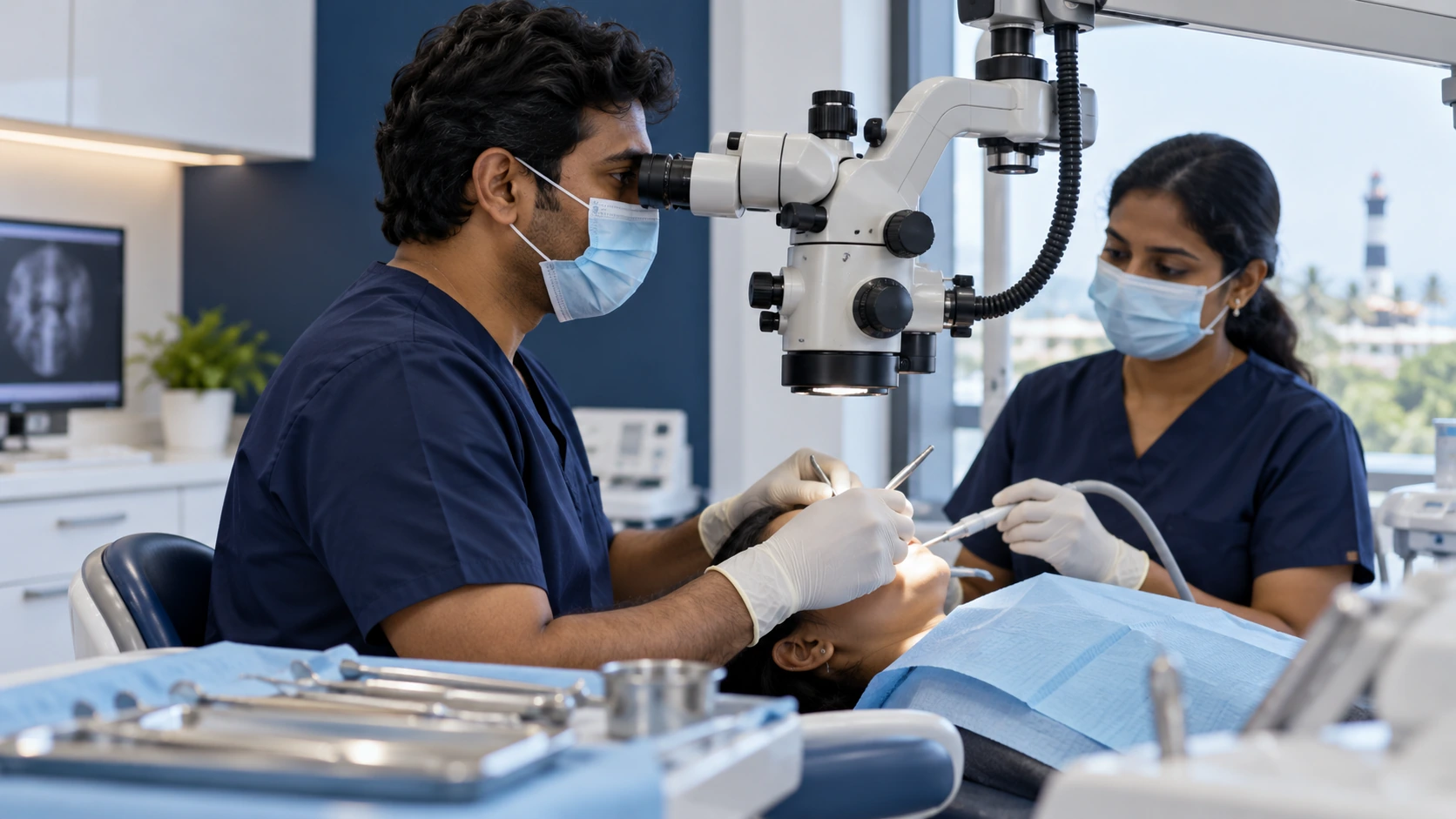
Microscope Dentistry in Pondicherry: Complete Guide to Precision Dental Care
Microscope dentistry is a modern approach that uses high-powered dental microscopes to provide enhanced magnification and illumination during selected dental procedures.
Unlike treatment performed only with the naked eye, a dental microscope can help the dentist visualize small and difficult-to-see structures in greater detail. This can be particularly useful for complex root canal treatment, restorative dentistry, diagnosis, and selected cosmetic procedures.
If you are searching for microscope dentistry in Pondicherry, understanding how dental microscopes work and where they may be useful can help you make an informed decision about your treatment.
Smile and Glow Dental Care provides microscope-assisted dentistry in Pondicherry as part of its advanced dental care services.
What Is Microscope Dentistry?
Microscope dentistry uses a specialized dental operating microscope to magnify the treatment area and provide enhanced illumination.
According to Smile and Glow Dental Care, its dental microscope can provide magnification of treatment areas up to 25 times their normal size. This enhanced visualization is used for selected root canal, restorative, and cosmetic procedures.
Microscope-assisted dentistry may help dentists identify fine details that are difficult to visualize with the naked eye.
How Does a Dental Microscope Work?
A dental operating microscope combines:
- High magnification
- Focused illumination
- Optical visualization
- Adjustable positioning
- Detailed visualization of small structures
The dentist views the treatment area through the microscope while performing the procedure.
The level of magnification can be adjusted according to the treatment requirement.
Why Is Magnification Important in Dentistry?
Teeth contain many small structures that can be difficult to see clearly.
Enhanced magnification may help dentists visualize:
- Small root canal openings
- Fine cracks
- Existing restorations
- Tooth anatomy
- Small areas of decay
- Treatment surfaces
- Fine details during restorative procedures
Better visualization can support more precise treatment in selected clinical situations.
Microscope Dentistry vs Treatment With the Naked Eye
Traditional dental treatment can be performed successfully using appropriate clinical examination, lighting, instruments, and magnification systems.
A dental microscope provides an additional level of magnification and illumination.
Conventional Visualization
Dentists may use:
- Dental operating lights
- Mirrors
- Dental instruments
- Loupes
- Clinical examination
Microscope-Assisted Visualization
A dental microscope can provide:
- Higher magnification
- Enhanced illumination
- Detailed visualization
- Better access to small structures
- Improved visibility during selected procedures
Microscope dentistry does not replace clinical skill or diagnosis. It is a tool that can support treatment where enhanced visualization is useful.
What Are the Benefits of Microscope Dentistry?
Potential benefits include:
- Enhanced visualization
- Better illumination
- Detailed examination
- Improved access to small structures
- Support for precise treatment
- Conservative treatment planning in selected cases
- Better documentation in some workflows
The actual benefits depend on the procedure, tooth condition, dentist's experience, and overall treatment plan.
Microscope Dentistry for Root Canal Treatment
Root canal treatment is one of the areas where magnification can be particularly useful.
Root canals can have complex anatomy, narrow openings, curves, and additional canals.
A dental microscope can help the dentist visualize:
- Canal openings
- Fine anatomical details
- Calcified areas
- Small cracks
- Existing filling material
- Complex canal structures
This can be especially relevant in difficult or previously treated root canal cases.
Why Use a Microscope for Root Canal Treatment?
Root canal treatment involves working inside very small spaces.
Enhanced magnification can help with:
- Locating canals
- Identifying complex anatomy
- Removing selected obstructions
- Evaluating cracks
- Improving visibility during cleaning
- Supporting precise restorative access
However, microscope use does not guarantee that every canal or complication can be identified or treated successfully.
The outcome depends on the tooth anatomy, diagnosis, infection, restoration, treatment technique, and long-term maintenance.
Microscope-Assisted Root Canal Treatment in Pondicherry
At Smile and Glow Dental Care, microscope-assisted dentistry is integrated into selected root canal procedures.
The clinic describes its microscope dentistry service as helping clinicians detect fine details and perform accurate, conservative treatment.
A root canal consultation may include:
- Clinical examination
- Dental X-rays
- Pulp testing when appropriate
- Magnified examination
- Treatment planning
- Root canal treatment
- Final restoration
The exact diagnostic and treatment process depends on the individual tooth.
Microscope Dentistry for Complex Root Canals
Some root canal cases are more complicated because of:
- Curved canals
- Narrow canals
- Calcification
- Unusual anatomy
- Multiple canals
- Previous root canal treatment
- Broken instruments
- Persistent infection
Magnification may provide useful visualization during selected complex procedures.
The dentist determines whether microscope-assisted treatment is appropriate after examination.
Microscope Dentistry for Root Canal Retreatment
A previously treated tooth may sometimes require root canal retreatment.
Possible problems can include:
- Missed canals
- Persistent infection
- Inadequate previous cleaning
- Broken instruments
- Leakage
- Complex canal anatomy
A microscope can provide enhanced visualization during selected retreatment procedures.
The dentist first needs to identify why the previous treatment did not achieve the desired result.
Microscope Dentistry for Detecting Cracks
Fine cracks can sometimes be difficult to identify.
Magnification and enhanced lighting can help dentists examine suspicious areas more closely.
A cracked tooth may present with:
- Pain while chewing
- Sensitivity
- Intermittent pain
- Unexplained discomfort
- Symptoms around an existing restoration
Microscope examination is only one part of diagnosis. The dentist may also use clinical tests, bite tests, imaging, and other diagnostic methods.
Microscope Dentistry for Restorative Treatment
Magnification can also be useful during selected restorative procedures.
It may help dentists work with greater visibility when:
- Removing decay
- Preparing teeth
- Evaluating margins
- Placing restorations
- Finishing restorations
- Examining existing dental work
The objective is to preserve healthy tooth structure where clinically appropriate.
Microscope Dentistry for Cosmetic Dentistry
Microscope-assisted visualization can also support selected cosmetic procedures.
It may be useful when evaluating:
- Fine tooth details
- Restoration margins
- Tooth surfaces
- Small defects
- Cosmetic restorations
Cosmetic treatment still requires comprehensive assessment of tooth shape, colour, alignment, gums, bite, and overall smile.
Microscope Dentistry for Dental Crowns
Dental crowns require accurate preparation and proper fit.
Magnification can assist in selected stages of:
- Tooth preparation
- Margin evaluation
- Restoration placement
- Finishing
- Bite assessment
Digital scanning and CAD/CAM workflows can also be combined with clinical magnification where appropriate.
Microscope Dentistry for Dental Fillings
Microscope-assisted restorative treatment may be useful for selected fillings, particularly when detailed visualization is needed.
The dentist may use magnification to:
- Identify small areas of decay
- Preserve healthy tooth structure
- Evaluate the treatment area
- Finish the restoration carefully
Not every routine filling requires microscope-assisted treatment.
Microscope Dentistry for Dental Diagnosis
Magnification can support a detailed visual examination of selected dental problems.
It may help evaluate:
- Fine cracks
- Restoration defects
- Tooth surfaces
- Root canal access areas
- Small anatomical structures
However, diagnosis should combine clinical examination with appropriate diagnostic tests rather than relying on magnification alone.
Who May Benefit From Microscope Dentistry?
Microscope-assisted treatment may be considered for patients with:
- Complex root canal anatomy
- Difficult root canal treatment
- Previous root canal treatment
- Suspected fine cracks
- Complex restorative requirements
- Difficult-to-visualize dental structures
- Selected cosmetic procedures
- Conservative restorative treatment needs
A dentist can determine whether microscope assistance adds clinical value to your specific treatment.
Is Microscope Dentistry Suitable for Everyone?
Not necessarily.
A dental microscope is a treatment tool rather than a separate treatment that every patient needs.
Routine dental procedures may not require high magnification.
The dentist may recommend microscope-assisted treatment when enhanced visualization can provide meaningful support for the procedure.
Is Microscope Dentistry Painful?
The microscope itself does not cause pain.
It is simply used to improve visualization during treatment.
Any discomfort depends on the dental procedure being performed.
For example:
- Dental examination is generally non-invasive.
- Root canal treatment may require local anaesthesia.
- Tooth preparation for a crown may require local anaesthesia.
- Dental surgery is an invasive procedure and requires appropriate pain-control measures.
Your dentist should explain the expected experience before treatment.
Does Microscope Dentistry Make Root Canal Treatment Better?
A microscope can provide enhanced visualization during selected root canal procedures.
This may be useful for identifying small anatomical details and improving visibility.
However, the success of root canal treatment depends on multiple factors, including:
- Correct diagnosis
- Tooth anatomy
- Infection
- Cleaning and shaping
- Filling and sealing
- Final restoration
- Oral hygiene
- Follow-up care
A microscope is an important tool in selected cases, but it is not a guarantee of treatment success.
Microscope Dentistry and Minimally Invasive Treatment
Enhanced visualization can support conservative treatment in selected cases.
By seeing the treatment area more clearly, the dentist may be able to work precisely while preserving appropriate healthy tooth structure.
The extent of tooth preservation depends on:
- Existing decay
- Tooth damage
- Previous treatment
- Cracks
- Tooth anatomy
- Restoration requirements
Microscope Dentistry for Dental Trauma
Dental trauma can cause:
- Tooth fractures
- Cracks
- Pulp damage
- Root damage
- Restoration failure
Magnification may assist in examining fine tooth details after trauma.
However, trauma assessment may also require X-rays and other diagnostic tests.
Microscope Dentistry for Previously Treated Teeth
Previously treated teeth can sometimes develop new symptoms or require additional treatment.
Magnification may assist during evaluation of:
- Existing root canal fillings
- Access cavities
- Canal anatomy
- Restoration margins
- Cracks
- Retreatment procedures
The dentist will determine whether retreatment, restoration, surgery, or another option is appropriate.
Microscope Dentistry Treatment Process
A typical microscope-assisted dental procedure may involve several stages.
Step 1: Consultation
The dentist discusses your symptoms, dental history, and concerns.
Step 2: Examination
The tooth and surrounding tissues are examined.
Step 3: Diagnostic Testing
Depending on the case, the dentist may use:
- X-rays
- Pulp testing
- Bite testing
- Clinical examination
- Digital imaging
Step 4: Treatment Planning
The dentist determines whether microscope assistance can provide a clinical advantage.
Step 5: Magnified Treatment
The dental microscope is positioned and adjusted according to the procedure.
Step 6: Treatment
The selected dental procedure is completed under enhanced visualization.
Step 7: Restoration
The tooth may require a filling, crown, or other restoration.
Step 8: Follow-Up
The dentist reviews healing and treatment progress when required.
How Long Does Microscope Dentistry Take?
The duration depends on the procedure.
Root Canal Treatment
Treatment time depends on:
- Number of canals
- Canal complexity
- Infection
- Tooth location
- Previous treatment
Restorative Treatment
Simple restorations may require less time than complex procedures.
Retreatment
Previously treated teeth may require more time because of existing materials and complex anatomy.
Comprehensive Treatment
Multiple procedures naturally require several appointments.
Your dentist can provide a more specific estimate after examination.
How Much Does Microscope Dentistry Cost in Pondicherry?
There is no single fixed cost for microscope dentistry.
The overall cost depends primarily on the underlying dental procedure.
Factors may include:
- Type of treatment
- Tooth location
- Root canal complexity
- Number of canals
- Previous treatment
- Dental materials
- Restoration required
- Laboratory work
- Additional diagnostic procedures
A consultation is required for an accurate treatment estimate.
Does Microscope Dentistry Cost More?
Microscope-assisted treatment may involve specialized equipment, clinical expertise, and additional treatment complexity.
However, the cost should be considered in relation to the complete dental procedure rather than treating microscope use as a standalone service.
The dentist can explain whether magnification is necessary for your case and how it affects the treatment plan.
Microscope Dentistry vs Dental Loupes
Both dental microscopes and dental loupes provide magnification.
Dental Loupes
Loupes are worn by the dentist and provide magnification directly through specialized lenses.
Dental Microscope
A dental microscope provides adjustable magnification and illumination and can offer a higher level of visualization for selected procedures.
The appropriate magnification system depends on the procedure and dentist's clinical approach.
Microscope Dentistry vs Digital Dentistry
Microscope dentistry and digital dentistry are different technologies that can complement each other.
Microscope Dentistry
Primarily improves:
- Magnification
- Visualization
- Illumination
- Precision during selected procedures
Digital Dentistry
Primarily supports:
- Digital scanning
- Imaging
- Treatment planning
- CAD/CAM restorations
- Digital implant planning
- Orthodontic planning
Both can be used together in comprehensive dental treatment.
Microscope Dentistry and Digital Imaging
Digital imaging can provide information about structures that cannot be seen directly.
Microscope visualization provides magnified views of accessible treatment areas.
Combining:
- Clinical examination
- Digital imaging
- Magnification
- Other diagnostic tests
can provide a more complete picture of selected dental problems.
Why Choose Smile and Glow Dental Care for Microscope Dentistry?
Smile and Glow Dental Care provides microscope-assisted dentistry in Pondicherry.
Its microscope dentistry service uses enhanced magnification and illumination for selected:
- Root canal treatments
- Restorative procedures
- Cosmetic procedures
- Complex dental treatments
The clinic states that its microscope-assisted approach is intended to improve visualization and support accurate, conservative treatment. :chatgpt-content-reference{index="1"}
Smile and Glow Dental Care also lists microscope dentistry alongside digital dentistry, root canal treatment, digitally designed crowns, implants, aligners, laser treatment, and other advanced dental services. :chatgpt-content-reference{index="2"}
Microscope Dentistry Consultation in Pondicherry
If you have persistent tooth pain, a difficult root canal, a previously treated tooth, or a suspected crack, a dental examination can help determine whether microscope-assisted treatment is appropriate.
During your consultation, the dentist may evaluate:
- Symptoms
- Tooth structure
- Root canal anatomy
- Existing restorations
- Previous treatment
- Gum condition
- Bite
- Dental imaging
The dentist can then explain whether magnification is useful and which treatment options are available.
How to Maintain Your Tooth After Microscope-Assisted Treatment
Microscope-assisted treatment does not remove the need for normal dental care.
After treatment:
- Brush twice daily
- Clean between your teeth
- Attend recommended follow-up appointments
- Avoid biting hard non-food objects
- Complete the recommended final restoration
- Maintain crowns and restorations
- Report persistent pain or swelling
- Attend regular dental checkups
Good oral hygiene and regular dental care remain important for long-term results.
Final Thoughts: Microscope Dentistry in Pondicherry
Microscope dentistry uses advanced magnification and illumination to help dentists visualize fine dental structures during selected procedures.
It can be particularly useful for:
- Complex root canal treatment
- Root canal retreatment
- Fine crack evaluation
- Restorative dentistry
- Conservative dental treatment
- Selected cosmetic procedures
A dental microscope is a clinical tool, not a replacement for diagnosis or professional expertise.
The most appropriate treatment begins with a detailed dental examination and diagnosis. If enhanced magnification can provide additional clinical value, microscope-assisted treatment may be incorporated into the treatment plan.
If you are looking for microscope dentistry in Pondicherry, a consultation can help determine whether this technology is suitable for your particular dental condition.
Frequently Asked Questions
What is microscope dentistry?
Microscope dentistry uses a high-powered dental operating microscope to provide enhanced magnification and illumination during selected dental procedures.
What is microscope dentistry used for?
It can be used for selected root canal treatments, root canal retreatment, restorative procedures, crack evaluation, and certain cosmetic dental procedures.
How much can a dental microscope magnify?
Smile and Glow Dental Care states that its dental microscope can magnify treatment areas up to 25 times their normal size, depending on the procedure and magnification setting.
Can microscope dentistry help with root canal treatment?
Yes. Enhanced magnification can help dentists visualize small canal openings, complex anatomy, fine cracks, and other details during selected root canal procedures.
Is microscope-assisted root canal treatment painful?
The microscope itself does not cause pain. Root canal treatment is generally performed using local anaesthesia, with the patient's comfort considered throughout the procedure.
Can a microscope detect a cracked tooth?
Magnification and enhanced illumination can help a dentist examine fine cracks more closely, but crack diagnosis may also require clinical tests and dental imaging.
Is microscope dentistry better than normal dentistry?
A microscope provides enhanced visualization, but it is not required for every dental procedure. Its value depends on the complexity and clinical requirements of the treatment.
Does microscope dentistry cost more in Pondicherry?
The cost depends primarily on the underlying dental treatment, its complexity, materials, restoration requirements, and other procedures involved. A consultation is required for an accurate estimate.
How long does microscope-assisted dental treatment take?
The duration depends on the procedure. Complex root canals and retreatments may require more time than routine restorative procedures.
Can microscope dentistry be used for root canal retreatment?
Yes. Enhanced visualization can be useful when examining previously treated teeth and performing selected retreatment procedures.
What is the difference between a dental microscope and dental loupes?
Both provide magnification. Loupes are worn by the dentist, while a dental operating microscope provides adjustable magnification and enhanced illumination for selected procedures.
Where can I get microscope dentistry in Pondicherry?
Smile and Glow Dental Care provides microscope-assisted dentistry in Puducherry for selected root canal, restorative, cosmetic, and complex dental procedures.