


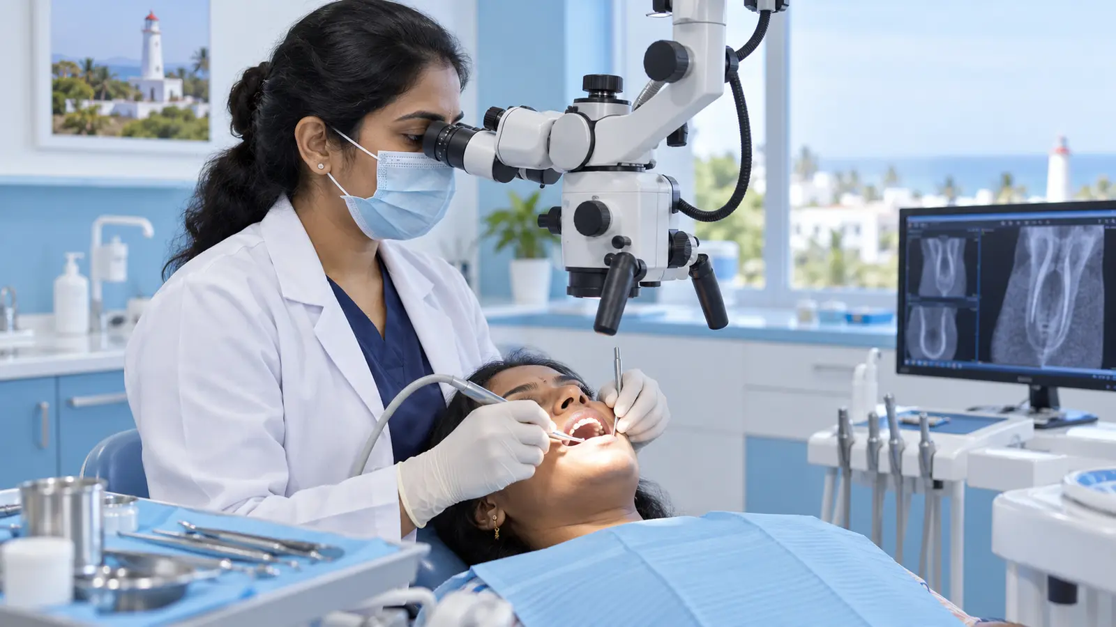
Microscope Dentistry for Root Canal Care in Pondicherry
A dental operating microscope provides strong illumination and magnification that can help a trained dentist identify fine anatomy, cracks, calcification or previous treatment materials during selected root canal procedures.
Key takeaways
- Magnification improves visibility and illumination.
- It can help with complex anatomy and retreatment.
- Diagnosis, isolation and disinfection remain essential.
- No technology can guarantee a treatment result.
Microscope Dentistry for Root Canal Care in Pondicherry: what patients should know
Root canal systems are small and can contain narrow branches, additional canals or changes from previous treatment. A dental microscope enlarges the working field and directs light into the tooth. It does not guarantee a particular outcome, but it can improve visual information and support precise, conservative work when used with sound diagnosis and technique.
How a dentist evaluates the concern
Magnification may be especially useful when canals are difficult to locate, a tooth has unusual anatomy, an instrument or post must be assessed, a previous root canal needs review, or a suspected crack requires closer inspection. The dentist still relies on clinical tests and radiographs; the microscope is one part of the diagnostic and treatment system.
Treatment planning and realistic choices
The patient experience is similar to other root canal appointments. The microscope remains outside the mouth while the clinician looks through it. Rubber dam isolation, irrigation, canal measurement and sealing remain essential. Digital imaging and ultrasonic instruments may be combined with magnification when clinically appropriate.
Care, recovery and long-term follow-up
Ask why magnification is recommended in your case and whether treatment will be performed by a clinician experienced with the equipment. After treatment, follow the same principles: protect the tooth, complete the final restoration, maintain oral hygiene and attend the advised review. Technology supports care but does not replace follow-up.
When to seek urgent help
Complexity should not delay assessment of severe symptoms. Facial swelling, fever, trauma or difficulty swallowing requires prompt attention. If a previous root canal has become painful or developed a gum swelling, take old X-rays or treatment records to the consultation when available.
Questions to take to your Pondicherry appointment
Ask the clinician to explain the diagnosis, which findings support it, reasonable alternatives, expected appointments, important risks, aftercare and the complete estimated treatment scope. Mention your medicines, allergies, pregnancy, medical conditions and previous dental treatment. This information helps the dental team plan safer, more personal care.
How to prepare for a useful consultation
Write down when the concern began, what makes it better or worse, and whether it affects eating, sleeping or daily activities. Bring recent dental records or radiographs when available, along with a current list of medicines, allergies and important medical conditions. Avoid starting antibiotics or pain medicines on someone else’s advice. A clear history, clinical examination and appropriate imaging help the dentist distinguish the likely cause from conditions that can appear similar.
Choosing care with confidence
A professional recommendation should be based on your examination rather than a fixed package or an online estimate. Ask what may happen if treatment is delayed, whether monitoring is reasonable, what alternatives exist, who will provide each stage and what follow-up is included. Patients travelling within or to Puducherry should also plan enough time for review visits. Treatment outcomes vary, so informed consent and realistic expectations are more valuable than promises of a guaranteed result.
Key points for informed dental care
- Magnification improves visibility and illumination.
- It can help with complex anatomy and retreatment.
- Diagnosis, isolation and disinfection remain essential.
- No technology can guarantee a treatment result.
SMILEKRAFT DENTAIRE provides patient-focused dental assessment in Puducherry. Explore the related dental service or request an appointment for an individual evaluation.
Frequently asked questions
Does the microscope go inside the mouth?
No. It is positioned above the patient while the clinician views the tooth through magnified optics.
Is microscope dentistry needed for every filling?
No. Its value depends on the procedure, anatomy and clinical need.
Can magnification find every crack?
It can improve visual inspection, but some cracks remain difficult to confirm and diagnosis may require several findings.
Does a microscope make root canal treatment painless?
Comfort is managed with anaesthesia and careful technique; magnification primarily supports visibility and precision.
Sources and further reading
Local context: Puducherry, Puducherry